lymphoma thyroid cancer ultrasound colors
Anatomic and physiologic assessment of the thyroid. Malignant lymph nodes in the neck whether they are metastatic from the thyroid or elsewhere ie.

Hashimoto S Thyroiditis And A Giraffe Pattern On Thyroid Ultrasound
Typical imaging findings include fine sand-like calcification cystic necrosis and.

. Objective Primary thyroid lymphoma PTL is an uncommon thyroid malignancy. Methods Twenty-seven pathologically confirmed PTLs were. J Ultrasound Med 34.
This study was designed to investigate the sonographic features of PTL. Despite the rarity of PTL it is important to recognize PTL promptly because its management differs from that of all the other thyroid neoplasms. Breast cancer advocates absolutely own the color pink.
This paper reviews the. Thyroid ultrasound reveals slight increase in size of lobes from 3 512x9 mm to 4121x 12mm and isthmus is1. It has gradually become widely accepted that this entity often represents primary involvement of the gland comparable to that seen in other extranodal sites 1.
Lymphoma usually occurs within lymph nodes but in rare cases it arises from lymphocytes that are present within the thyroid gland. The sonographic findings for 13 surgically proven primary thyroid lymphomas were analyzed and compared to those for 27 nodular goiters. In accordance with the suggested.
Biopsy confirmed non-Hodgkin lymphoma of the thyroid gland. Various ultrasound findings in patients with a thyroid mass. Thyroid nodule ultrasound color doppler.
Coloring pages for adults free teens carcinoma prostate ultrasound and color imaging a gallery of high resolution images thyroid cancer colors ca. That said in general the following colors are associated with the following cancer types and it should be noted that while some colors and associations make sense others do not and arent meant to. Lymphoma is a cancer of the white blood cells that can be found in the blood stream or in lymph nodes.
12 patients 80 had a solitary nodule type 1 two 13 had multiple nodules type 2 and one 7 had a diffuse goiter type 3. It is not unusual for this type of cancer to involve the surrounding structures such as the trachea or esophagus. The clinical presentation of lymphoma as a thyroid mass is rare.
Objectives The purpose of this study was to correlate the clinicoradiologic and pathologic features of thyroid lymphoma and to identify the most useful diagnostic method for thyroid lymphoma as the. Secondary thyroid lymphoma affects lymph nodes and other organs first followed by subsequent spread to the thyroid. Microcalcifications were found in 38 of cancerous nodules and only in 5 of benign non-cancerous nodules.
The clinical picture described in several series is distinct and uniform but treatment results have been variable 15. This could not be mistaken for any other cancer. The CT appearances were classified into three types.
The risk of cancer increased with the size of. Staging is a tool your doctor uses to classify characteristics. Most often it is not detected until it gets to a certain size that would make it physically prominent.
Other links to color or gray scale. Color doppler ultrasound also plays a vital role in the diagnosis of thyroid cancer. Both lymphoma and thyroid cancer are quite common individually lymphoma of the thyroid gland is also frequently found while their joint presentation is more unusual.
Thyroid lymphoma typically progress rapidly creating a large mass in the thyroid which often causes compressive symptoms difficult breathing difficulty swallowing or voice changes. Abnormal thyroid cancer ultrasound colors friday february 18 2022 edit. This is called primary thyroid lymphoma to distinguish it from lymphomatous.
Burkitt lymphoma of thyroid gland which is a very rare subtype. Thyroid nodules were found in 97 of patients with thyroid cancer and in 56 of without thyroid cancer. Lymphoma is a cancer that develops in the lymphatic system the tissues and organs that produce store and carry white blood cells.
Cronan JJ Scola FH. Lymphoma thyroid cancer ultrasound colors july 24 2021 july 24 2021 0 comment 1206 am method of brunn is widely used to calculate thyroid volume tvol as a sum of the two lobes each calculated according to the following formula. On average 1 case of thyroid cancer was found for every 111 ultrasound exams performed.
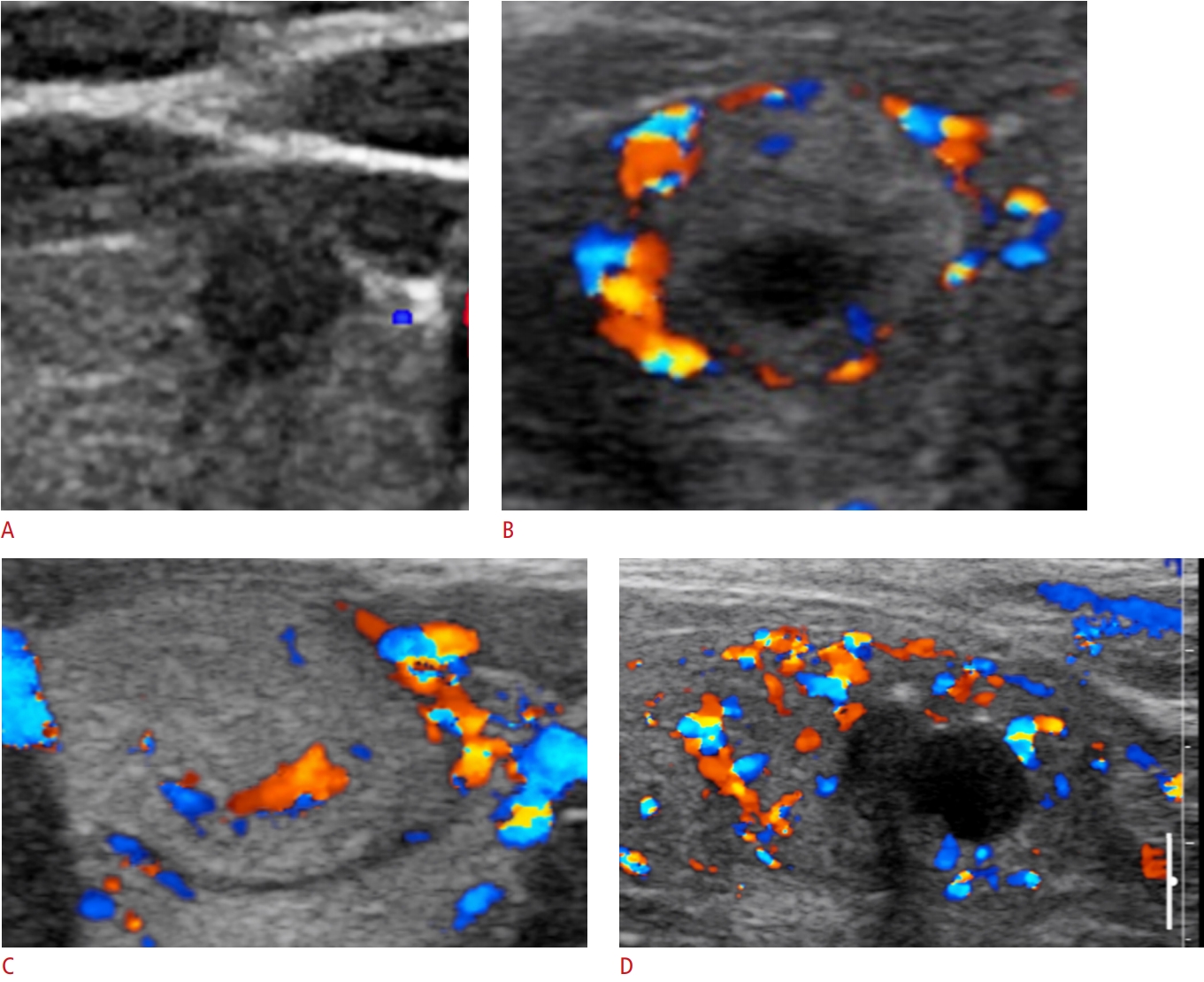
In A the two-part figure on the left shows a thyroid adenoma with a peripheral halo sign arrows in a nodule that is very well circumscribedThe companion color flow image shows a predominately peripheral pattern of flow suggestive of benign disease. The purpose of this study was to determine the specific sonographic features of primary thyroid lymphoma and its color Doppler pattern compared to nodular goiter. The use of ultrasound in thyroid cancer imaging is dealt with in more detail in the later part of the review but in brief its major roles include.
The appearance of primary thyroid lymphoma on computed tomographic CT scans and clinical data for 15 patients were analyzed. Ultrasound features of anaplastic carcinoma include fig.

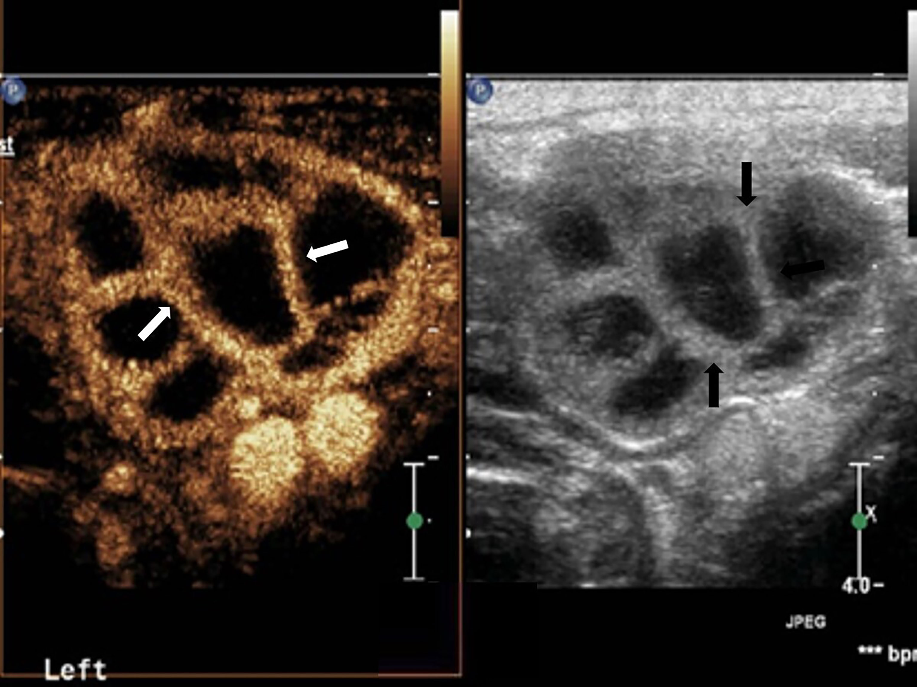
Frontiers Reassessing The Value Of Contrast Enhanced Ultrasonography In Differential Diagnosis Of Cervical Tuberculous Lymphadenitis And Lymph Node Metastasis Of Papillary Thyroid Carcinoma

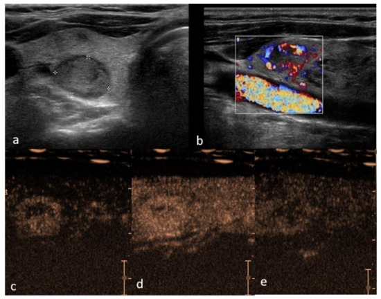
Cancers Free Full Text Performance Of Contrast Enhanced Ultrasound In Thyroid Nodules Review Of Current State And Future Perspectives Html

Ultrasound Of A 42 Year Old Man Who Incidentally Detected A Thyroid Download Scientific Diagram

Mesenteric Adenitis Medical Ultrasound Ultrasound Sonography Pelvic Inflammatory Disease

Subacute Thyroiditis An Often Overlooked Sonographic Diagnosis Lee 2016 Journal Of Ultrasound In Medicine Wiley Online Library
5 Best Ways To Diagnose Thyroid Cancer

Reports Free Full Text Ultrasonographic Findings In Common Thyroid And Parathyroid Disorders Advantages Of Real Time Observation By The Endocrinologist With Their Own Ultrasound Machine Html

Primary Thyroid Lymphoma Has Different Sonographic And Color Doppler Features Compared To Nodular Goiter Wang 2015 Journal Of Ultrasound In Medicine Wiley Online Library

Ultrasound Features Of The Various Types Of Thyroid Lesions Panel A Download Scientific Diagram

Imaging And Screening Of Thyroid Cancer Radiologic Clinics

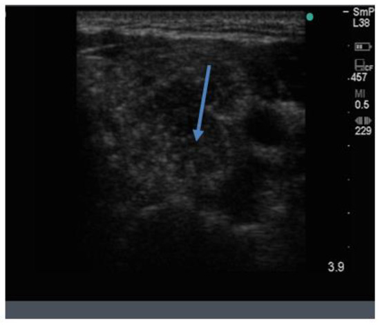
A Thyroid Ultrasound Shows A Markedly Hypoechoic Area Arrow With Download Scientific Diagram

Papillary Necrosis Ultrasound Google Search Medical Ultrasound Ultrasound Ultrasound Sonography

Thyroid Doppler Ultrasound Us Showing Hypo Download Scientific Diagram

Primary Thyroid Lymphoma Has Different Sonographic And Color Doppler Features Compared To Nodular Goiter Wang 2015 Journal Of Ultrasound In Medicine Wiley Online Library


